QUESTION-1:
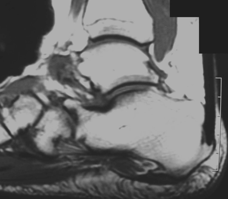
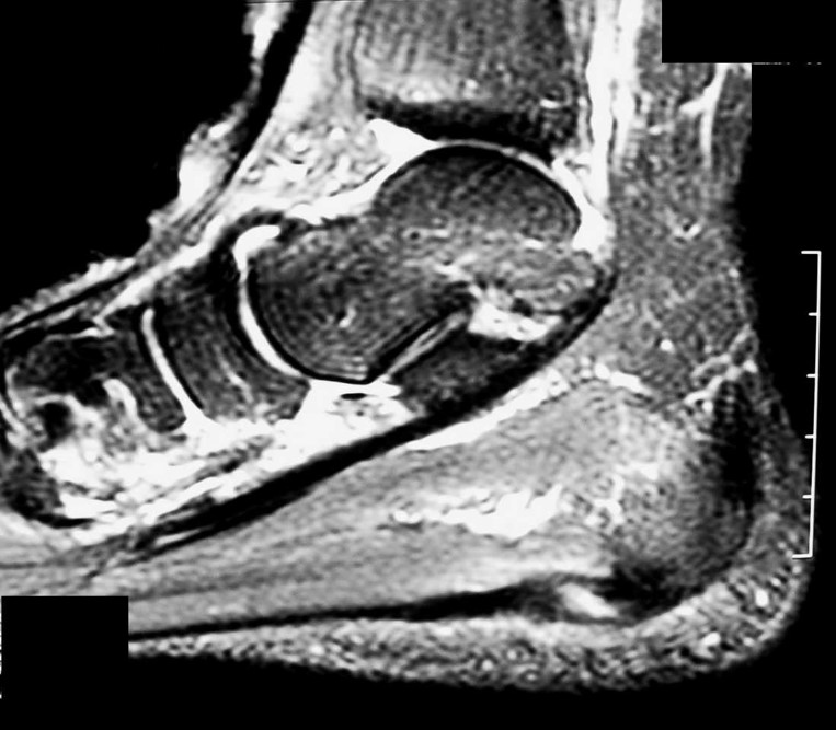
63 YEAR OLD FEMALE WITH FOOT PAIN:
Correct Answer: Plantar Fasciitis
Differential Diagnosis:
- Calcaneal Stress Fracture
- Fascial Rupture
- FHL Tendinitis
- Tarsal tunnel syndrome
Pearls:
- Principal complaint : Pain on under surface of heel after weight bearing.
- Pain can be worse when weight is borne after a period of rest (e.g. in morning)
- Eases with walking
- Passive dorsiflexion of the toes may exacerbate discomfort.
- mechanical: stress of repetitive trauma (more common)
- degenerative
- systemic: as an enthesopathy in association with seronegative spondyloarthropathies
- Ankylosing spondylitis
- Reiter Syndrome
- Psoriatic arthritis
Clinical presentation:
Pathology:
Generally a low-grade inflammatory process involving the plantar aponeurosis with or without the involvement of the perifascial structures. It can arise from several factors:References:
- https://radiopaedia.org/articles/plantar-fasciitis?lang=gb


QUESTION-2:
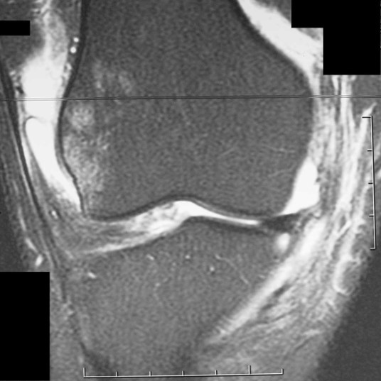
15 YEAR OLD SOCCER PLAYER:
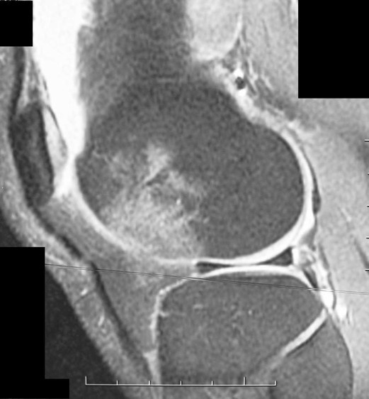
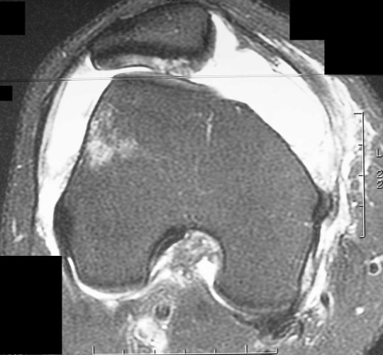
Correct Answer: Transient Patellar dislocation
Differential Diagnosis:
- Acute ACL tear
- Patellar subluxation
- Trochlear dysplasia
Pearls:
- Occurrence : Patellar dislocation accounts for 2-3% of all knee injuries and is commonly seen in those individuals who participate in sports activities.
- Attempt to slow forward motion while pivoting medially on a planted foot internal rotation of femur and quadriceps contraction produces a net lateral force.
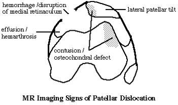
Mechanism:
References:
- https://radiopaedia.org/articles/lateral-patellar-dislocation?lang=gb


QUESTION-3:
26 YEAR OLD FEMALE STATUS POST FALL:
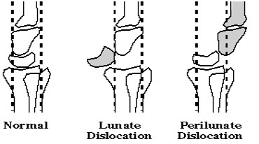
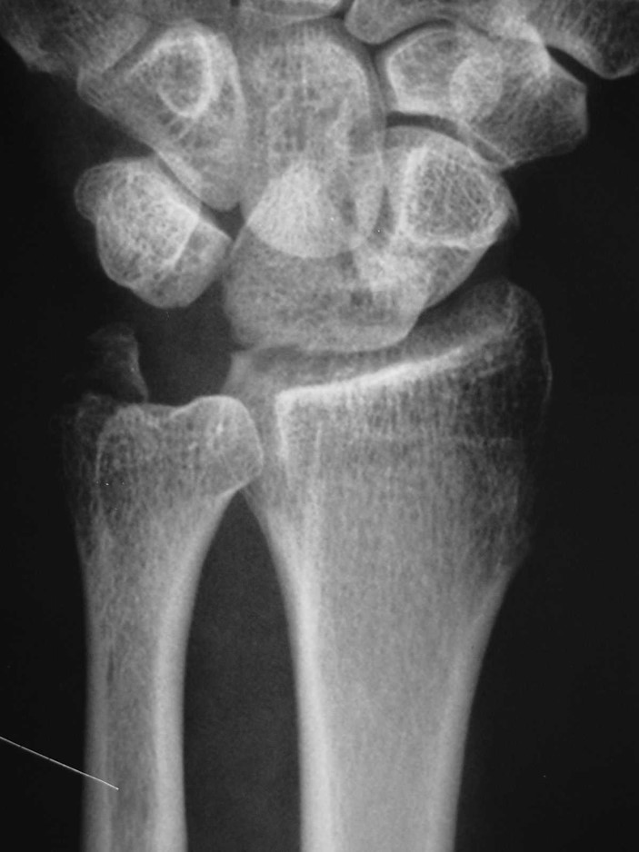
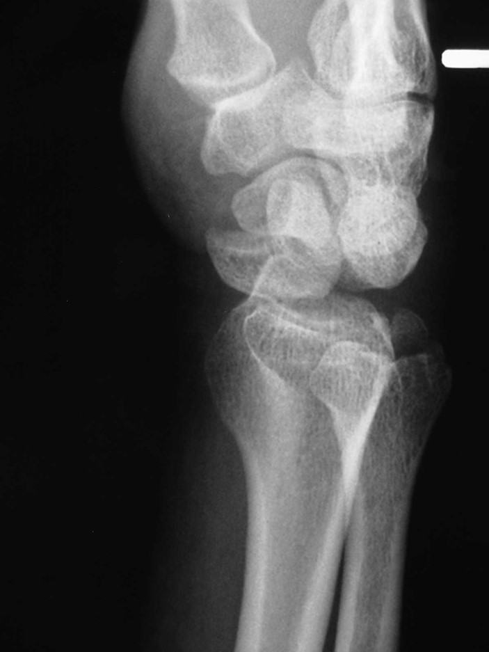
Correct Answer: Lunate dislocation
Differential Diagnosis:
- Peri lunate dislocation
- Midcarpal dislocation
- Triquetral fracture
Pearls:
- Fall on outstretched hand
- Ligament b/t Lunate & Capitate Disrupted
- Sometimes difficult to differentiate from peri lunate
- For surgeons, moot point as just want rapid reduction of C-L dislocation
- Failure to recognize can result in median nerve damage by lunate impingement
Mechanism:
References:
- https://radiopaedia.org/articles/lunate-dislocation#:~:text=Lunate%20dislocations%20are%20an%20uncommon,in%20relation%20to%20the%20radius.


QUESTION-4:
69 year old black male:
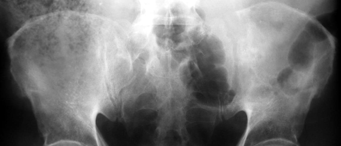
Correct Answer: Plasmacytoma
Differential Diagnosis:
- Metastasis
- Chondrosarcoma
- Chronic osteomyelitis
- Osteoblastoma
Pearls:
- Absent systemic involvement
- < 5% plasma cells in routine blind bone marrow biopsies
- Very low serum monoclonal immunoglobulin levels
Imaging:
- Possible cortical disruption and soft tissue extension
- Lytic, well circumscribed, and often expansile
- Otherwise normal survey
- Distribution = MM
- MRI role: screening of the spine and pelvis reveals radiographically unsuspected lesions
Features:
References:
- https://radiopaedia.org/articles/plasmacytoma


QUESTION-5:
42 year old female with fluctuant mass:
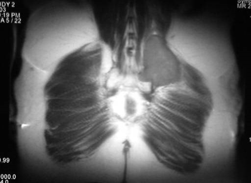
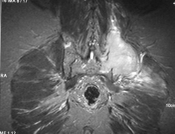
Correct Answer: Lipoma
Differential Diagnosis:
- Angiolipoma
- Myolipoma
- Chondroid Lipoma
- Lipoblastoma
- Spindle cell lipoma
- Hibernoma
Pearls:
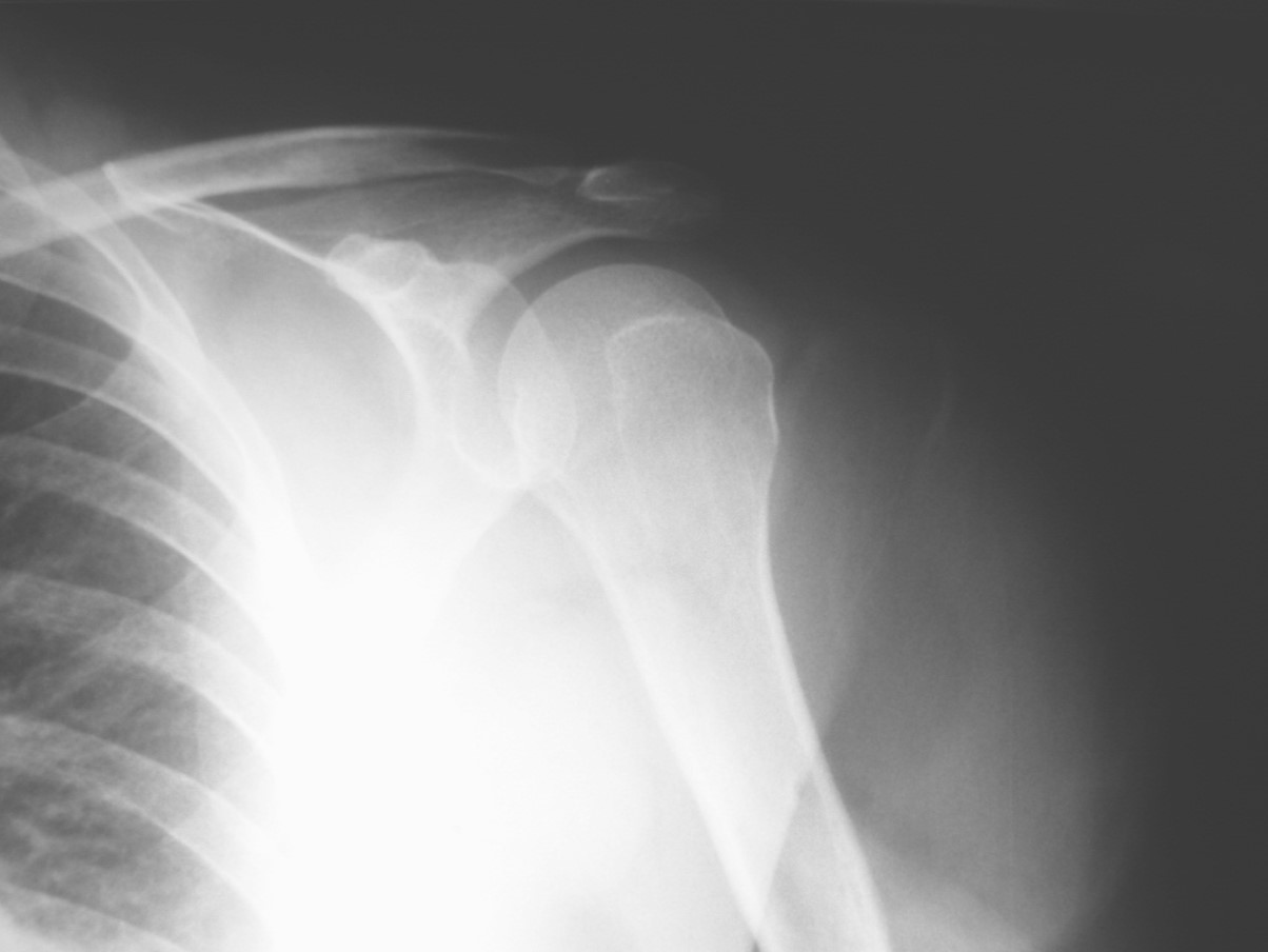
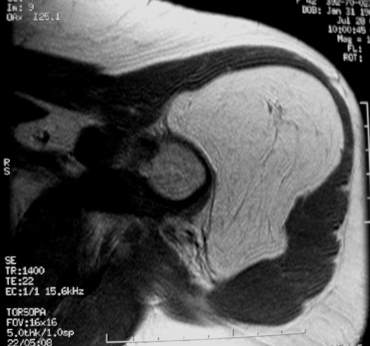
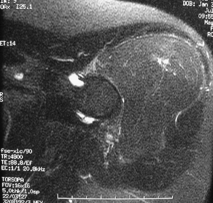
- Most common soft tissue tumor
- Mature adipose
- Age 50-70
- Common Locations:
- Deep: Chest Wall, Retroperitoneum, Hands, Feet
- Superficial: Back, Neck, Shoulder, Abdomen, Gluteal
- Multiple 5-8%
- May grow, but usually stabilize
- MRI
- Fat Intensity on all Sequences
- No enhancement
- May have fibrous septa
- Rx: Local excision or Observe
References:
- https://radiopaedia.org/articles/lipoma


QUESTION-6:
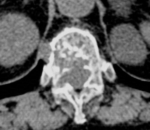
60 yo afebrile with acute back pain:
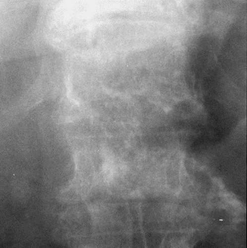
Correct Answer: Chordoma
- Chordoma is a destructive bone tumor believed to arise from notochord cell rests, which are normally precursors of vertebrae and intervertebral discs. The usual clinical presentation is in males between the ages of 40 and 70 years. It is locally malignant with a strong tendency to recur after attempted excision. Metastatic spread is unusual. The lesions are slow-growing and become apparent due to pressure symptoms, with or without localized pain. Approximately half of the lesions arise in the sacrum and/or coccyx, the others in the basioccipital and basisphenoid regions of the skull.
- A vertebral origin is found in only 15% of patients. Adjacent vertebrae may be involved.
- When the lesion is located in the sacrum, plasmacytoma and giant cell tumor should be considered in the differential diagnosis. When the lesion is located in the basioccipital region, chondrosarcoma should be considered. Because of the possibility of the presence of chordoma in vertebral bodies, we suggest that it should be included in the differential diagnosis of osteolytic lesions of this area.
- The other 2 choices are typically Sclerotic or blastic bone diseases.
Differential Diagnosis:
- EXPANSILE LESION IN THE POSTERIOR ELEMENTS
- Giant cell tumor
- Osteoblastoma
- ABC
- Plasmacytoma
References:
- https://bit.ly/3rMdUZh


QUESTION-7:
Why is a CT scan of the hip performed after reduction of a dislocated hip?:
Correct Answer: Evaluate for fracture fragment in the hip joint
Posterior Hip Dislocation – 80 to 85%; associated with fractures of the posterior rim of the acetabulum, femoral head anterior Hip Dislocation – 5 to 10%; associated with fractures of the acetabular rim, greater trochanter, femoral neck, and femoral head. MRI may be useful to detect osteonecrosis of the femoral head. Sources: Dähnert 5th p. 67; Greenspan 3rd p. 221-222


QUESTION-8:
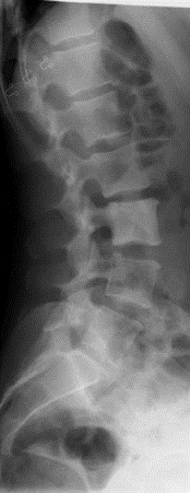
65 yo male with known malignancy:
Correct Answer: Lymphoma
- Solitary dense vertebrae, as well as in lymphoma, are also seen in osteoblastic metastases (prostate/breast), hemangioma (coarse trabecular pattern, vertical in orientation).
Differential Diagnosis:
- IVORY VERTEBRAL BODY
- Paget’s
- Osteopetrosis
- Mastocytosis
- Mets - Lymphoma
- Sarcoidosis
- SSDz
Pearls:
- Paget's disease (tends to involve the posterior elements as well as displaying expansion of the body), osteopetrosis, sickle cell anemia, fluorosis, systemic mastocytosis, tuberculous infection, metastatic carcinoid tumor, osteoblastoma, osteosarcoma, and even primary Ewing's sarcoma are other considerations.


QUESTION-9:
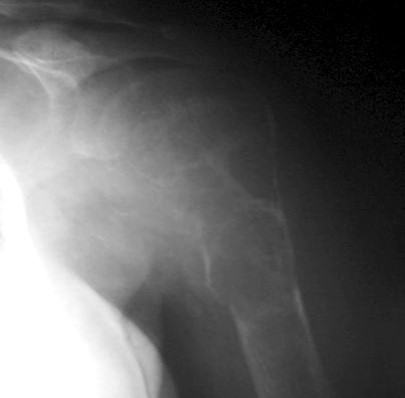
52 yo patient with chronic shoulder pain:
Correct Answer: Brown Tumor
- The commonest lesion showing fluid-fluid levels with hemorrhage on CT and/or MRI is primary or secondary ABC. The presence of fluid-fluid levels in an osseous lesion is not pathognomonic of a specific lesion.
The association of fluid-fluid levels and brown tumors has recently been reported. Although not previously reported, it is not unexpected to find fluid-fluid levels in brown tumors since these tumors often contain hemorrhage.
Langerhans cell histiocytosis: primarily occurs in older children and young adults, with a male to female ratio of 2:1.
Differential Diagnosis:
- Aneurysmal bone
- Telangiectatic osteosarcoma
- Chondroblastoma(occurring predominantly in young patients (<20 years of age))
- Giant cell tumor of bone.
Fluid-fluid levels have commonly been reported to occur
References:
- https://bit.ly/3C4He1y



QUESTION-10:
An Epileptic presents with frozen shoulder. Most likely cause:
Correct Answer: Posterior dislocation
- 2-4% of shoulder dislocations. Traumatic causes include convulsive disorders and electric shock therapy; nontraumatic causes voluntary, involuntary, congenital, developmental.Types: subacromial, subglenoid, subspinousIn > 50% unrecognized initially + subsequently misdiagnosed as frozen shoulder; average inury between injury & diagnosis is 1 year. Sources: Dähnert 5th p. 68; Greenspan 3rd p. 111


QUESTION-11:
An Epileptic presents with frozen shoulder. Most likely cause:
Correct Answer: Posterior dislocation
- 2-4% of shoulder dislocations. Traumatic causes include convulsive disorders and electric shock therapy; nontraumatic causes are voluntary, involuntary, congenital, developmental. Types: subacromial, subglenoid, subspinous > 50% unrecognized initially + subsequently misdiagnosed as a frozen shoulder; average injury between injury & diagnosis is 1 year. Sources: Dähnert 5th p. 68; Greenspan 3rd p. 111.


QUESTION-12:
What inserts on the lesser tuberosity?:
Correct Answer: Subscapularis
- The supraspinatus, infraspinatus, and teres minor insert on the greater tuberosity.Source: Greenspan p. 91


QUESTION-13:
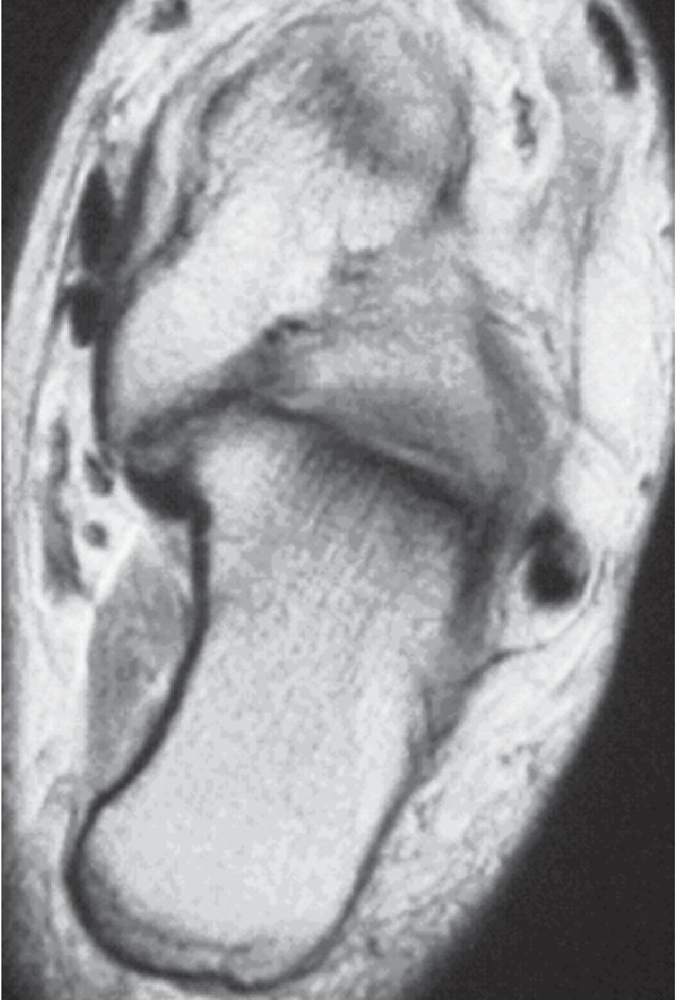
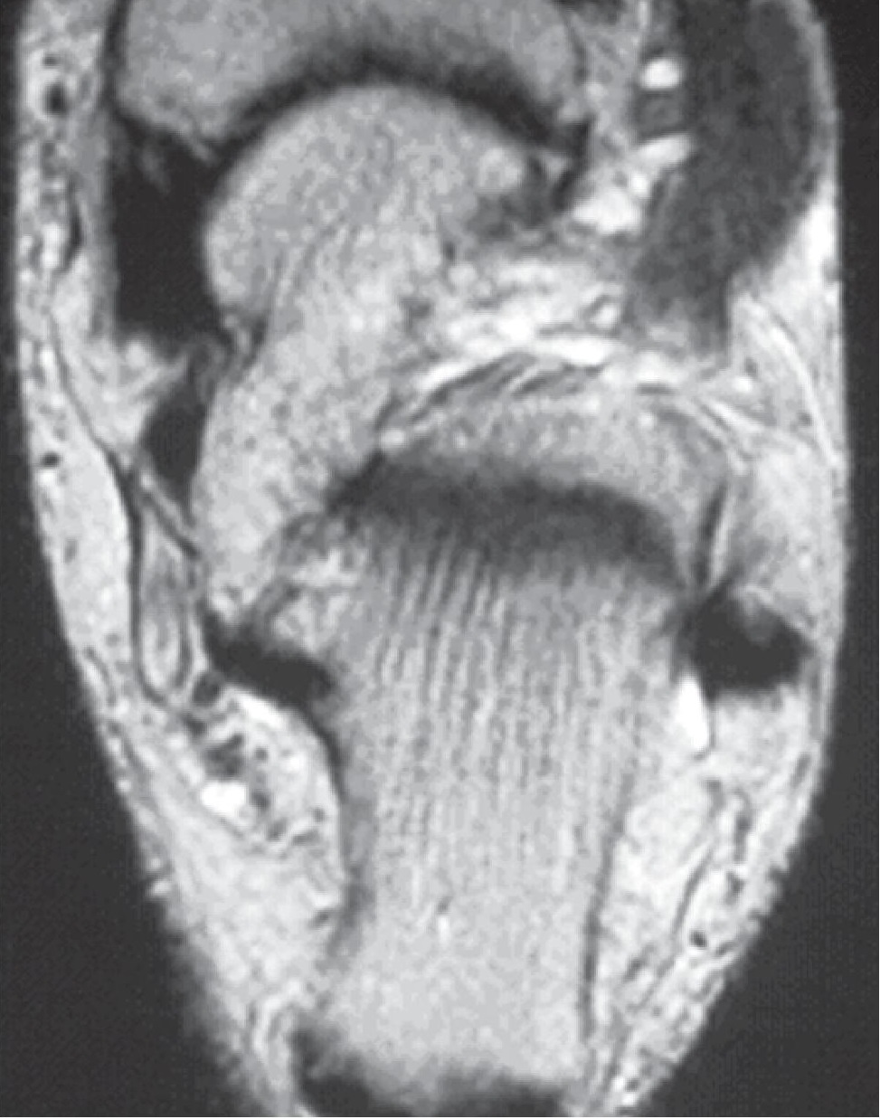
You are shown axial T1- and T2-weighted MR images (Figures 1A and 1B) of a 23-year-old woman with foot pain. Which one of the following is the MOST likely diagnosis?:
Correct Answer: Subtalar coalition
- A. Incorrect. Posterior tibial tendon tears may be incomplete with thickening of the tendon, type I, incomplete with tendon attenuation, type II and complete, type III. Associated fluid at the tendon sheath is typical. Prominence of the medial soft tissues may be present. The posterior tibial tendon in the test case is normal.
- B. Correct. The medial facet of the subtalar joint is one of the most common sites of developmental coalition of the foot. Such unions may be fibrous, cartilaginous or bony. In either case, there is close approximation of the 2 bones, often with down-sloping of the sustentaculum tali as seen in the test case.
- C. Incorrect. Dysplasia epiphysealis hemimelica—also known as Trevor’s disease, tarsal aclasis, tarsoepiphyseal aclasis—is a development anomaly (dysplasia) in which a bony outgrowth, exostosis like in appearance, develops at an epiphysis or epiphysioid bone (epiphysealis) usually involving one side of the joint and one side of the body (hemimelica). The most common sites are the ankle and knee. The abnormal appearance of the medial subtalar joint in the test case represents coalition, not a bony outgrowth.
- D. Incorrect. Tarsal tunnel syndrome is a compressive neuropathy involving the posterior tibial nerve resulting from any space-occupying lesion, behind and below the medial malleolus, beneath the flexor retinaculum. The tarsal tunnel in the test case is normal.
Differential Diagnosis:
- Narrowing with closely opposed irregular articular interface at the medial subtalar joint with maldevelopment of the sustentaculum tali, which slopes downward.
References:
- Stoller, Tirman, Bredella. Diagnostic Imaging Orthopaedics. Amirsys Inc. Salt Lake City, UT.
- Resnick, Niwayama. Diagnosis of Bone and Joint Disorders. W.B. Saunders. Philadelphia, PA. Fourth Ed.


QUESTION-14:
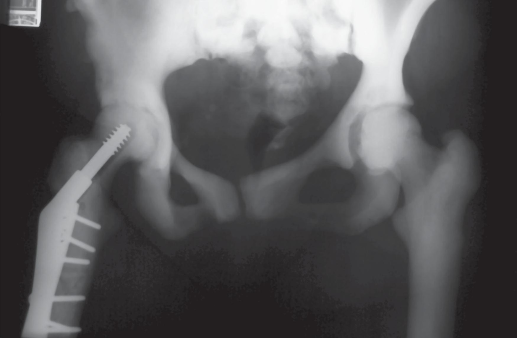
You are shown an AP view (Figure 2) of the pelvis and hips of a 44-year-old woman. What is the MOST likely diagnosis?:
Correct Answer: Osteopetrosis
- A. Correct. Osteopetrosis is a sclerosing dysplasia involving a defect in osteoclastic resorption of the primary spongiosa related to endochondral bone formation. Sclerosis is usually diffuse and uniform. Loss of the cortical medullary junction is typical. Alternating bands of sclerosis may also be present reflecting the periodicity of the disease. In the adult form or delayed type, autosomal dominant variety, originally described by Albert-Schoenberg, the patient’s life expectancy may be normal but the skeleton is brittle and prone to fracture.
- B. Incorrect. Although the pelvis is a common site for osteoblastic deposits in patients with tuberous sclerosis, they are usually multiple and discrete rather than diffuse and uniform as in the test case.
- C. Incorrect. Renal osteodystrophy does not produce such uniform widespread sclerosis with loss of the cortical medullary junction as in the test case. When there is extensive involvement, sclerosis tends to be more patchy and ill-defined related to secondary hyperparathyroidism.
- D. Incorrect. The sclerosis of sickle cell disease is secondary to infarction and osteonecrosis which over time is replaced by fibrosis and new bone formation. The diffuse, uniform sclerosis at the pelvis and hips with diffuse loss of the cortical-medullary junction would be unusual.
Differential Diagnosis:
- Diffuse uniform sclerosis of the pelvis and hips with failure of differentiation between cortex and medullary cavity, status post hip fixation.
References:
- Greenspan. Orthopaedic Radiology. Lipincott Williams Wilkins. Philadelphia, PA. Third Ed.
- Resnick, Niwayama. Diagnosis of Bone and Joint Disorders. W.B. Saunders. Philadelphia, PA. Fourth Ed


QUESTION-15:
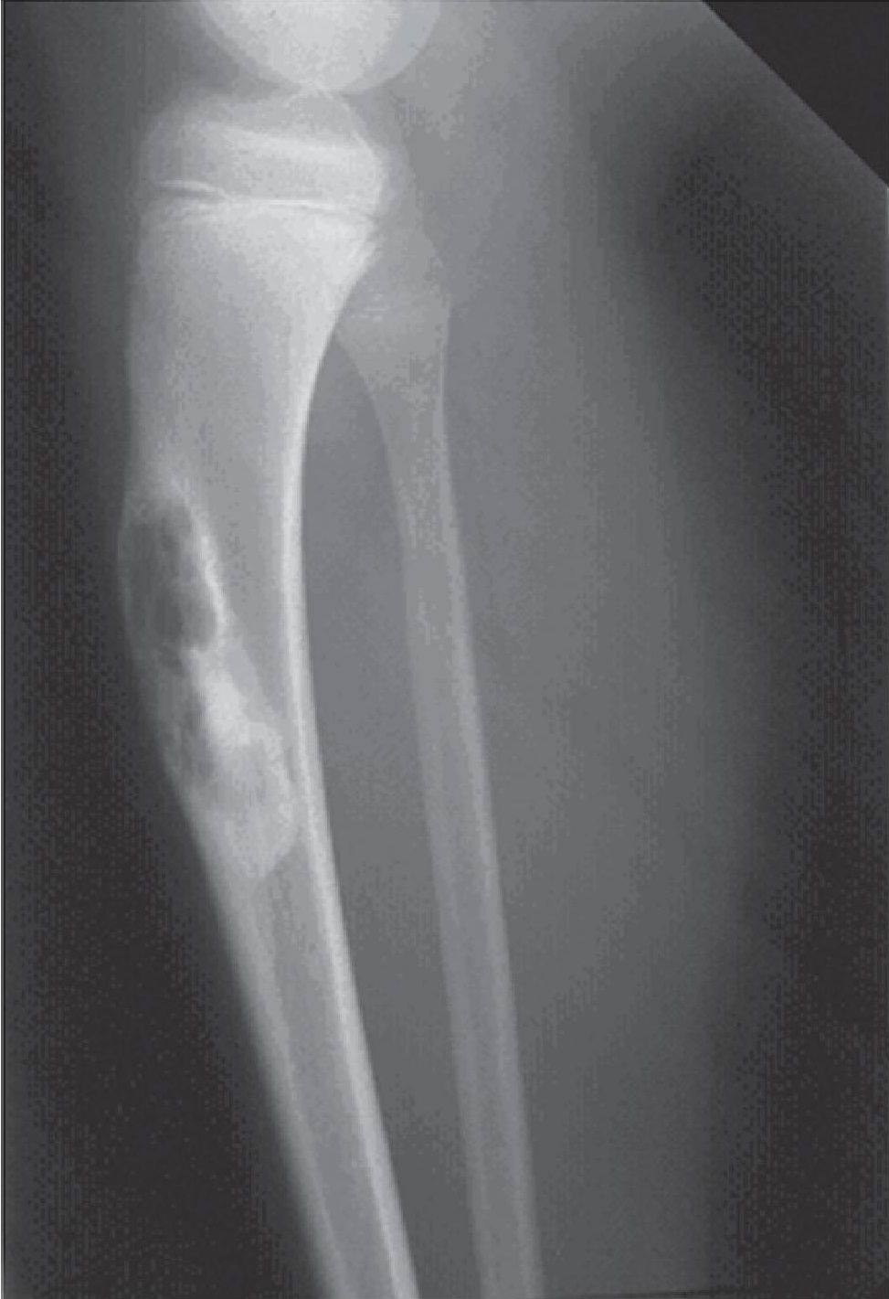
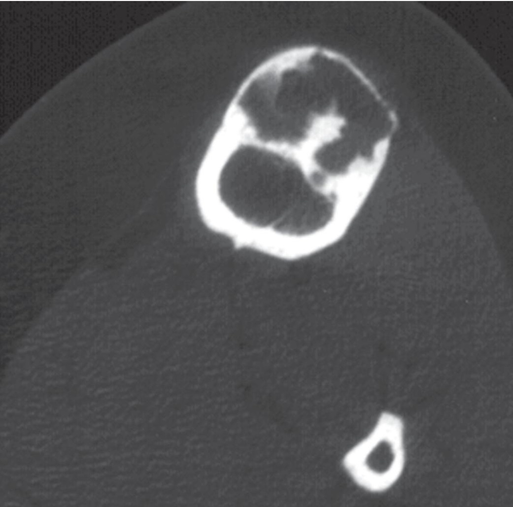
You are shown a lateral radiograph (Figure 3A) and a non-contrast CT (Figure 3B) of a 9-year-old boy with anterior leg bowing. What is the MOST likely diagnosis?:
Correct Answer: Osteofibrous dysplasia
- A. Incorrect. Intracortical osteosarcoma is the most uncommon form of osteosarcoma. They are diaphyseal, usually arising in the femur or tibia. Intracortical osteosarcoma generally presents as lucency within the cortex that measures less than 4 cm, with surrounding sclerosis. If small, they may be mistaken for an osteoid osteoma or fibrous cortical defect.
- B. Incorrect. Nonossifying fibromas may be mildly expansile, but they are not associated with bowing of the bone.
- C. Correct. Osteofibrous dysplasia is a benign fibro-osseous lesion that is almost exclusively found in the tibia or fibula. It is a disorder of childhood, usually seen within the first 2 decades of life and often under 10 years of age. The lesion is centered in the anterior cortex, and may be associated with anterior bowing. A lobulated lucency is seen, often with surrounding sclerosis. The appearance may mimic a nonossifying fibroma, but the location and bow are key. Adamantinoma may be present in association with osteofibrous dysplasia. The two disorders cannot be differentiated one from the other by imaging.
- D. Incorrect. Aneurysmal bone cysts are eccentric, lytic lesions that are usually found in the medullary cavity. They are most often metaphyseal, but may be present anywhere along a bone. Cortical and periosteal locations have been reported, but are unusual. The cortex overlying the lesion is expanded and if growth is rapid, it may be destroyed.
Differential Diagnosis:
- There is a cortically based, expansile lesion involving the anterior tibia shaft.
- The tibia is mildly bowed anteriorly.
- The majority of the lesion is lucent, with a lobulated contour and sclerotic margins.
References:
- Huvos AG. Bone Tumors: Diagnosis, Treatment, and Prognosis, 2nd Ed. WB Saunders Company, Philadelphia, PA, 1991.
- Unni KK, Ed. Dahlin’s Bone Tumors: General Aspects and Data on 11, 087 cases, 5th ed. Lippincott-Raven Publishers, Philadelphia, PA, 1996


QUESTION-16:
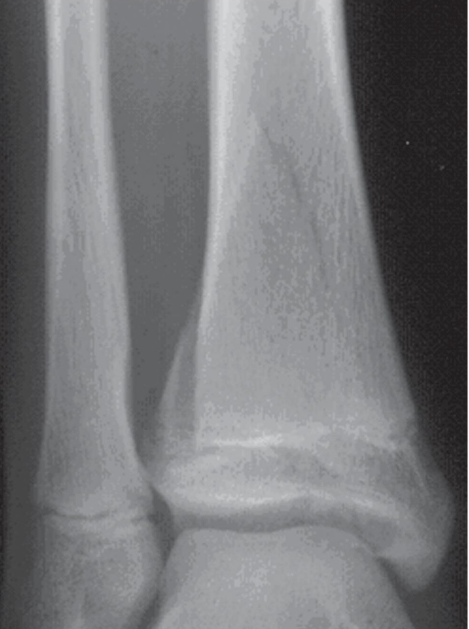
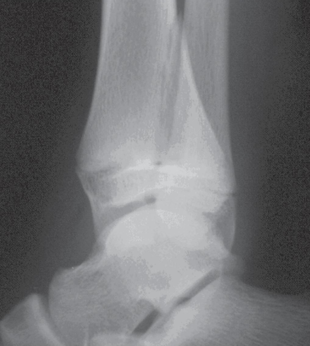
You are shown frontal and lateral radiographs (Figures 4A and 4B) of a12-year-old boy who fell from his skateboard. What is the fracture type?:
Correct Answer: Triplane
- A. Incorrect. A Tillaux fracture is a Salter type III avulsion of the anterolateral aspect of the distal tibial epiphysis.
- B. Correct. The radiograph shows a coronally oriented fracture of the distal metaphysis of the tibia, a horizontally oriented fracture of the lateral aspect of the physis, and a sagittally oriented fracture of the distal epiphysis, all of which comprise a triplane fracture. The injury results in part due to the partially fused tibial physis, closure progressing medial to lateral.
- C. Incorrect. A pilon fracture is a comminuted fracture of the distal tibia and tibial plafond due to impaction of the talus into the tibia.
- D. Incorrect. A Maisonneuve fracture involves the proximal fibula shaft and is associated with rupture of the distal syndesmotic ligaments and widening of the tibia-fibular syndesmosis.
Differential Diagnosis:
- A Salter IV fracture with a sagittal epiphyseal, transverse physeal and coronal metaphyseal orientation.
References:
- Greenspan. Orthopedic Radiology. A Practical Approach. 2nd ed. Gower Medical Publishing, NY, NY 1992.


QUESTION-17:
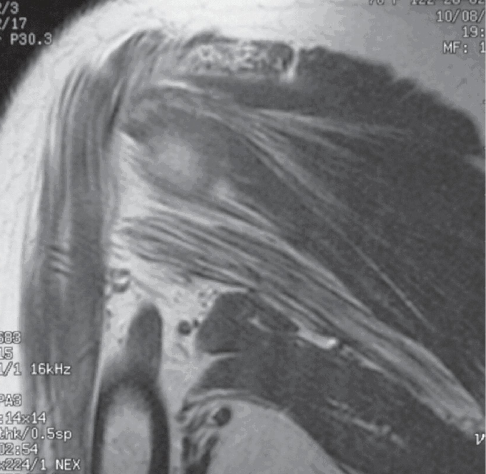
You are shown an oblique coronal T1-weighted MR image (Figure 5) of a 45-year-old woman with chronic mild shoulder pain. What is the MOST likely diagnosis?:
Correct Answer: Quadrilateral space syndrome
- A. Incorrect. Parsonage-Turner syndrome (also known as acute brachial neuritis) is an idiopathic denervation of the shoulder muscles resulting in pain and weakness. The muscles demonstrate high SI on T2-weighted imaging due to acute denervation edema and ultimately atrophy and fatty replacement. The suprascapular nerve and, therefore, the supraspinatus and infraspinatus musculature are typically involved. Axillary nerve involvement may occur but is less frequent.
- B. Incorrect. While chronic tears of the rotator cuff tendons may be associated with muscle atrophy and fatty replacement, no tears are demonstrated in the test case.
- C. Correct. Quadrilateral space syndrome is caused by compression of the axillary nerve in the quadrilateral space, usually by small fibrous bands or ganglion cysts and clinically manifests as mild shoulder pain. Axillary nerve dysfunction may eventually lead to atrophy of the teres minor and deltoid musculature.
- D. Incorrect. The teres minor and deltoid muscles are supplied by the axillary nerve, not the suprascapular nerve. The suprascapular nerve innervates the supraspinatus and infraspinatus muscles, which appear normal.
Differential Diagnosis:
- Atrophy teres minor and deltoid musculature.
References:
- Kaplan, Helms, Dussault, Anderson, Major. Musculoskeletal MRI. WB Saunders Co., Philadelphia, PA. 2001

QUESTION-18:
The radiographic appearance of SI joint widening is associated with ALL of the following EXCEPT:
Correct Answer: Hyperthyroidism
- A. Incorrect. Septic sacroiliitis may significantly erode the articular surfaces of the sacrum and ilium, producing enough bone loss to create an appearance of joint widening.
- B. Correct. Osteoporosis is the most characteristic feature of hyperthyroidism in the adult skeleton. In the immature skeleton, there may be acceleration of skeletal maturity. Thyroid acropachy is an unusual manifestation of thyroid disease usually observed after the treatment of hyperthyroidism at which time the patient may be hypothyroid, euthyroid or even hyperthyroid. Periosteal new bone formation at the small bones of the hands and feet is the characteristic radiographic finding. S-I joint involvement is not seen.
- C. Incorrect. Trauma to the pelvis may result in diastasis at one or both SI joints producing a radiographic appearance of SI joint widening.
- D. Incorrect. Secondary hyperparathyroidism is characteristic of renal osteodystrophy and may result in subchondral bone resorption at the SI joints.
References:
- Resnick, Niwayama. Diagnosis of Bone and Joint Disorders. W B Saunders, Philadelphia, PA Fourth Ed.

